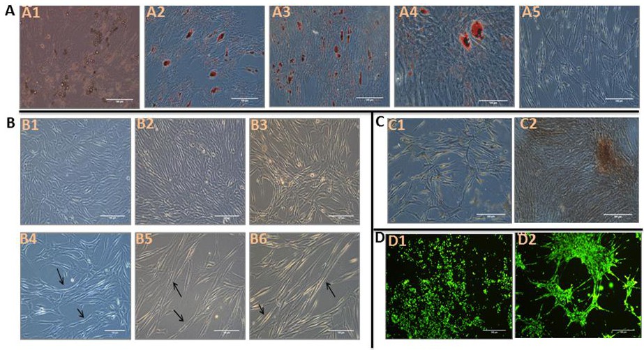
Fig. 3. Inverted microscope images showing multipotent differentiation potential of pericytes. A. Images of the adipogenic differentiation of pericytes on day 17 A1. shows the formation of lipid droplets in response to adipogenic induction before staining, (A2 and 3) showing differentiated pericytes on day 17 after staining with Oil red O, A4. magnified image of A3, A5. control pericytes that were not subjected to adipogenic induction but were stained with Oil red O. B. Images showing the myogenic differentiation of pericytes using 5-azacytidine, (B1-B3) control pericytes that not subjected to myogenic induction; black arrows indicate elongated myoblast-like cells on days 7,13 and 14. C. Images showing the osteogenic differentiation of pericytes on day 17. C1. images of Alizarin red-stained control cells that were not induced to undergo osteogenic differentiation. C2. showing Alizarin red-stained cells after osteogenic induction. D. Angiogenic differentiation (tube formation assay) of pericytes captured using an inverted fluorescence microscope. D1. control cells stained with Calcein AM without angiogenic induction. D2. cells stained with Calcein AM after angiogenic induction.